
byThe University of Hong Kong
Credit:Cell Biomaterials(2025). DOI: 10.1016/j.celbio.2025.100213
A research team from the Department of Orthopedics and Traumatology, School of Clinical Medicine, LKS Faculty of Medicine at the University of Hong Kong (HKUMed), has developed a titanium implant surface that can be activated by near-infrared (NIR). With just 15 minutes of NIR irradiation, this surface can eliminate 99.94% of Staphylococcus aureus (S. aureus) biofilms without the use of antibiotics, while simultaneously promoting bone-implant fusion.
Based ontitanium dioxide(TiO2), the same compound found in titanium's natural surface oxide layer, the design may offer practical advantages for compatibility with existing titanium implants and future clinical translation. This innovative technology could be applied to various common orthopedic implants, including joint replacements, fracture fixation devices, spinal fusion cages, dental implants and craniofacial implants. It offers a new solution to combat implant infections. The findings werepublishedas a cover story in the journalCell Biomaterials.
Implant-associated infections remain a major clinical challenge in orthopedic practice. Once bacteria adhere to an implant surface and formbiofilms, they become highly tolerant to antibiotics and can evade the patient's immune system. Consequently, patients often require repeated invasive debridement, revision surgeries and prolonged courses of high-dose systemic antibiotics. These treatments extend recovery time, increase health care costs, contribute to antibiotic resistance and often fail to prevent recurrent infections.
Professor Kelvin Yeung Wai-kwok, from the Department of Orthopedics and Traumatology, School of Clinical Medicine, HKUMed, who led the research, stated, "Implant-associated infection is a major cause of implant failure. Bacterial colonization and biofilm formation on implant surfaces can be difficult to eradicate, often resulting in persistent inflammation, compromised implant fixation and, ultimately, loosening and failure."
"Althoughexisting antibacterial coatingscan provide some level of protection, they typically rely on loaded agents such as antibiotics, metal ions or complex bioactive molecules," he added. "Their effectiveness, however, is limited by their loading capacity, uncontrolled release, potential cytotoxicity and loss of function once these agents are exhausted.
"These limitations underscore the urgent need for an in situ, controllable and durable strategy that can both eliminate biofilms and promote bone-to-implant integration without the use of additional drugs or agents. This is precisely what our research seeks to achieve."
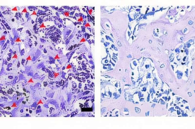
In a rat tibial implant model, on day 14 after surgery, the conventional titanium (upper left) exhibited obvious exudate and pus, indicating severe local infection and inflammation. In contrast, the hydrogenated nano-honeycomb (upper right) showed no obvious exudate or pus. Stained tissue sections further revealed numerous bacteria (red spots in images) in the conventional titanium at day 14, while almost no bacteria were detectable in the hydrogenated nano-honeycomb, highlighting its superior antibacterial performance in vivo. Credit:Cell Biomaterials(2025). DOI: 10.1016/j.celbio.2025.100213
Light-triggered defense enables rapid bacterial elimination
Currently, titanium and titanium alloys, which are widely used in orthopedic surgery, naturally form a very thin TiO2layer on the implant surface. While this layer is highly biocompatible, it offers minimal defense against infection and lacks the ability to actively promote bone growth. When bacteria such as S. aureus form biofilms on these surfaces, antibiotics are often unable to eradicate the infection effectively, resulting in the need for invasive revision surgeries.
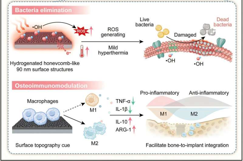
Using a customized template-assisted technique, the HKUMed team precisely engineerednano-honeycomb structureson the titanium surfaces and introduced oxygen vacancies through hydrogenation treatment, creating a remotely activatable smart surface. Under NIR irradiation, this smart surface generates reactive oxygen species and a mild local photothermal effect, rapidly disrupting biofilm architecture and killing bacteria.
In vitro experiments found that a single 15-minute irradiation was sufficient to eliminate 99.94% of S. aureus. In a rat tibial defect infection model, the same treatment removed 91.58% of biofilms. Compared to unmodified titanium implants, the engineered surface can efficiently perform biofilm clearance and lead to marked reductions in pus formation and local inflammation, resulting in a notable increase in new bone formation around the implant.
Enhancing the osteoimmune microenvironment supports new bone formation
Beyond its strong antibacterial performance, the research demonstrated that the new implant surface effectively modulates the local immune response. It shiftsmacrophagesfrom a prolonged pro-inflammatory state to a pro-healing, tissue remodeling phenotype, thereby creating a more favorable osteoimmune microenvironment. This attracts more osteogenic cells and facilitates their differentiation, leading to a significant increase in new bone formation.
As a result, the implant achieves faster and more stable integration with the bone, demonstrating that the new technology prevents infection while accelerating bone fusion.
"By adopting an agent-free strategy, our newly developed smart surface demonstrates remarkable antibacterial and pro-osteogenic capabilities without the use of additional drugs," explained Professor Yeung.
"This technology offers high translational potential, as it relies solely on native implant materials and mature manufacturing processes. This will facilitate regulatory approval, support scalable manufacturing and pave the way for future clinical adoption. We believe this innovation will broadly improve orthopedic surgical outcomes and benefit more patients."
More information Yizhou Zhu et al, NIR-responsive hydrogenated TiO 2 nanoscale honeycomb surface pattern for rapid S. aureus biofilm elimination and enhanced osteogenesis, Cell Biomaterials (2025). DOI: 10.1016/j.celbio.2025.100213


Post comments